Extraction of Stem Cells from Dental Pulp and Alveolar Bone
Thasonporn Termthong, M. Sc. (Periodontology)*
Viniramol Sriwattana, Diploma of Oral Surgery and Maxillofacial, Dental Council**
Thanyapat Vanichanon, M. Sc. (Medical Science)***
Matthana Jongka, M. Sc. (Biology)***
Nasikarn Muangklom, High Vocational School Certificate, Computer Business***
** Specialized Dentists, Dental Work Group, Priest Hospital, Department of Medical Service, MoPH ***Research Colleagues, Medical Genetics Research Center, Rajanukul Institute, Department of Mental Health, MoPH
Introduction
Stem cells are any cells that possess 2 characteristics: cells that can self-replicate and can differentiate into unique or diverse specialized cells when properly stimulated. One of the two cells in the division process will remain the same as a stem cell that is not transformed into specialized cell types. Stem cells found in human body after birth and adult stem cells can be stimulated to renew into other cells containing the same germ layer, for instance, bone marrow stem cells originated from embryo tissue of mesoderm which can replicate into myelo supportive stroma, muscle cell, bone cell and adipose cell, or stem cells in dental pulp stemmed from embryo tissue of neuron crest which can differentiate into dentinoblast, fibroblast or adipose cell. Adult stem cells are found in various tissues such as bone marrow - derived mesenchymal stem cells, adipose - derived stem cells, peripheral blood - derived stem cells, skin-derived stem cells, and cardiac stem cells, etc. Tooth is one source of stem cells found in the dental pulp stem cells; DPSCs and stem cells from human exfoliated deciduous teeth ;SHED, periodontal ligament stem cells ; PDLSCs, stem cells from apical part; SCAP or from dental follicle stem cells; DFSC2 and in the spongy bone of alveolar bone containing marrows which also lie stem cells.3
Nowadays, stem cells from human teeth have been utilized in various dental cares: organ tissue engineering to replace facial and oral organs such as mandibular condyle reconstruction2, tooth organogenesis4, 5, 6, 7, tissue engineering to replace partially damaged organs- periodontal complex comprising cementum, periodontal ligament and parts of alveolar bone8, 9, bone engineering for oral bone culture10 and mucosa for tissue culture in the mouth 11, engineering of dentin/pulp tissue12, dentin regeneration13, periodontal ligament regeneration14 and reinforced application of stem cells for pseudo root by stem cells superficial coating 15, etc.
The unpainful or unharmful ways of obtaining stem cells can easily be made during the natural shaking or falling-off periods of deciduous teeth. However, acquiring stem cells from the pulled tooth for the purpose of getting cured in the older people without deciduous tooth is required, for example, the third molar which is impacted or tilted to the cheek or minor molar is pulled out for orthodontics including bone ridge or projecting dental alveolus that needs to be cut off when wearing denture. These stem cells are derived from disposed organs. Papatpong Sirikururat3 reported that stem cells could be differentiated from alveolus bone on the palate but none of reports showed the separation from torus mandibularis.
Therefore, the research team focused on the cell extraction from torus mandibularis by comparing to stem cells derived from the dental pulp2,12,13,16,17 which was extensively reported. This was the very first trial of Rajanukul Institute. This could be of great benefits for the separation of stem cells in torus mandibularis of the individuals with intellectual disabilities accordingly.
Materials and Methods
The descriptive research of ex vivo study that had passed the approval of the Research Ethics Committees of Rajanukul Institute no. Lor Wor 31/07/51 was used in this study.
Research participants were 7 healthy persons who had no congenital diseases required for any dental treatments by pulling out the third molar or minor molar for the sake of orthodontics from Rajanukul Institute and Priest Hospital and 3 persons who needed treatment with cutting the projecting alveolar bone for the purpose of wearing denture from Priest Hospital. Then anti-biotics, steroids and/or immunosuppressants were given within 3 months prior to conducting the research.
Stem Cell Differentiation from Dental Pulp Tissues
Out growth method16 was applied by taking the third molar or the whole minor molar that was pulled out from alveolar socket to be scraped for tissue with surgical blade no#15, then applied povidoiodine for 2 minutes and 70% ethanol for 2 minutes to get rid of bacteria. Later, cemento- enamel junction was operated with sterile high speed diamond fissure bur. Then, filtered water was sprayed to reduce the heat around the notch around the tooth17 and washed it with phosphate buffer saline: PBS 2 times and MEM, Dulbeccos Modified Eagle Medium high glucose; Invitrogen: Gibco 1 time. Put the sharpless tool at the notch and twisted to break apart the tooth at the notch area. After that, the endodontic spoon was used to get the tissue of dental pulp into 35mm culture dish (Corning Incorporated) Sliced with sterile blade into small pieces of the sizes of 1x1x1 ml, placed on the culture dish with the same interval and put the culture media consisting of 10% fetal bovine serum (Invitrogen: Gibco) 1% antibiotic-antimycotic solution containing 10,000 unit of penicillin (base), 10,000 g of streptomycin (base) and 25 g of amphotericin B/ml utilizing Penicillin G (sodium salt) streptomycin sulfate, amphotericin B in 0.85% saline (Invitrogen: Gibco) 1% L - Glutamine; Glutamax-I supplement (Invitrogen: Gibco) in DMEM solution. Next, put the culture dish into incubator at 37 C with 5% carbondioxide and optimal relative humidity. The culture media was changed day after day.
Separation of Stem Cells from Alveolar Bone
Out growth method16 was used to separate stem cells from the targeted alveolar bone, out growth bone of mandible as an example. When periostium was opened, out growth bone was removed from alveolus with fissure carbide bur, using low speed straight hand piece under the sprayed coolant of normal saline solution. Then sterile forcep was used to take the cut pieces of the bones into 35 mm culture dish (Corning Incorporated). Sliced them with sterile blade into small pieces of the sizes of 1x1x1 ml, placed on the culture dish with the same interval and put the culture media with the same content with those of dental pulp method except for DMEM solution using 20% fetal bobine serum. Put the culture dish into incubator at 37 C with 5% carbondioxide and optimal relative humidity. The culture media was changed day after day.
Cell Culture
After 2-3 days cuboidal shape and spindle shape of cells started to grow from the tissue of dental pulp and piece of bone. Observed for 1-2 weeks whether cells had covered the 35mm culture dish. Then observed whether cells were not too crowded and transferred them into 60mm culture dish18 (Trypsinization). Old culture media was removed and 0.5 EDTA; ethylene diamine tetra acetic acid was added to cover all cells. Then culture dish was placed into the incubator at 37C for 5 minutes. The culture dish was brought to check with a microscope. Drained EDTA solution, flushed with proper cell media to remove cells from culture dish when cells began to shrink to form a circular shape. After that, drained 1 mm of floating media, placed the culture dish into the incubator at 37C with 5% carbondioxide and highest relative humidity. Next day spindle shape of cells were found on the surface, added 4 mm media and changed media day after day. Transferred cells into a new 60 mm culture dish and observed whether the dish was filled with cells and not too crowded.
Identification of stem cells could be made in 2 ways: Immuno-fluorescence to see STRO-1 marker and reverse transcription polymerase chain reaction (RT-PCR) to see CD29 and CD44.
Immuno-fluorescence Smear
Prepared cells with an autoclaved cover slip (Menzel-Glaser: German), then, placed into 12-well plate (Corning: USA), washed with de-ionized water (DI) 2 times and 1 time with phosphate buffer saline.
Sew sorted cells into cover slip with the density of 70,000 cells per ml. Cultured until cells till the whole slide was covered and not too crowded. Then, fixed cells with phosphate buffer saline 2 times and applied cold methanol. It was left for10 minutes. After that, washed with phosphate buffer saline 2 times and 1mm phosphate buffer saline was added. Kept at 4C until smeared (not over than 1 month).17,18
Smearing process: Took 2 thin glassess of cells from 12-well plate, one was for control and the other was for the test. 10% fetal bovine surum in phosphate buffer saline was dropped into the 2 thin glassess throuroghly. Left them for 30 minutes then dropped 100 l of 1 antiboy (mouse anti stro-1 monoclonal antibody MAB 4315: Chemicon international USA.) in 10% phosphate buffer saline with the ratio of density of 1:100 over the test slide while the same quantity of 10% phosphate buffer saline was added into control slide and left it in the dark area for 24 hours. After that, 2 thin glasses were washed with phosphate buffer saline on the shaker for 3 minutes 3 times. Dropped 100 l of 2ํ antibody (Goat anti mouse IgG Biotin conjugated affinity purified antibody: AP124B: Chemicon international USA) in10% phosphate buffer saline with the ratio of density of 1:500 over the 2 thin glassess and leave them in the dark area at room temperature for 40 minutes. Then washed the thin glassess with phosphate buffer saline on the shaker for 3 times for 5 minutes each time. Dropped 100 l of Strep FITC ; Goat anti mouse IgG Biotin conjugated affinity purified antibody; S3762; Sigma Aldrich in10% phosphate buffer saline with the ratio of density of 1:500 over the 2 thin glassess and leave them in the dark area at room temperature for 40 minutes. Then washed the thin glassess with phosphate buffer saline on the shaker for 5 minutes 3 times. Dropped 10 l of DAPI working solution with the ratio of density of 1:1000 into the thin glass over the cells, placed it up side down on the glass slide covered with a foil ready for testing for Stro-1 marker by 100x fluorescent microscope (Applied spectral imaging: USA). No green fluorescent light appeared on the control glass while the testing one displayed green fluorescent light on the cytoplasm of stem cells.19
3 spots of cells scanned through 100x microscope with Stro-1 display were randomly selected for the calculation of the proportion of cells that displayed Stro-1 marker to the total cells,20 photographed under DAPI and FITC mode and photos were overlapped to be photographed in order to count the numbers of cells that showed Stro-1 and total cells with Adobe Photoshop. ) STRO-1 marker and reverse transcription polymerase chain reaction (RT-PCR) to see CD29 and CD44 were also observed.
Cells were photographed, transferred, spun by 1200 rpm centrifuge for 10 minutes and measured for light intensity by spectrophotometer21 before RNA was extracted with RNA extraction kit (Qiagen).22 Complementary DNA (cDNA) was synthesized from the extracted RNA with super script III First -Strand Synthesis System for RT PCR (Invirogen)23 prior to examination of stem cells marker, CD29 and CD44 with Duplex PCR using CD29 primer, CD44 primer and mRNA GAPDH.
CD29 primer (Proligo: Sigma; Singapore) had the length of 346 pairs (base pair: bp) with melting point at 64C for both forward and reverse strings.
CD29 primer (Proligo:Sigma; Singapore) had the length of 231 pairs with the melting point of forward string and reverse string at 61 and 64C respectively.
mRNA GAPDH primer (Techdragon ; Hongkong) had the length of 84 pairs with the melting point of forward string and reverse string at 65.1 and 56.9C respectively.
With the mixture of the following substances to make the total volume of 25 l and place in PCR machine (thermal cycler) with the temperature and time set as follows:
Step 1 Set temperature at 95C for 15 minutes
Step 2 Set temperature at 95C for 1 minute
Step 3 Set temperature at 55C for 1 minute
Step 4 Set temperature at 72C for 1 minute
Step 5 Repeat Step 2-4 for 34 rounds
Step 6 Set temperature at 72C for 10 minutes
Step 7 Set temperature at 4C for 1 minute
PCR product was obtained when these processes were completed. DNA isolation was then conducted with Agarose gel electrophoresis24,25 by weighing 0.600g of agarose powder and 30l of 1xTris Borate EDTA (TBE) and placing them into 100l flasks. Put them into a microwave to melt for 1 minute, observe whether agarose solution was homogeneously clear. Dropped 5l out of 10mg/ml of ethidium bromide into cooling down agarose solution, shook well and poured it into a tray of gel with plastic combs stuck on it for 1 hour to let the gel solidated. Removed the plastic combs and put the tray on the electro- phoretic chamber (Amersham)] with TBE solution (1xTBE) over the gel. Add 100 bp marker solution mixted with 5 l bromophenol blue (Sigma B8026) into the first well. Put 5 l of PCR product in each well mixed with 2 l bromophenol blue except for the last well that filled with 100 pairs of marker solution mixed with 2 l bromophenol blue. Covered the chamber, turn on the electrical generator (Electrophoretic power supply: Amersham) with 69 volt, 2 ampere for 90 minutes. Then, placed the tray of gel into the ultraviolet chamber (Gel Doc: Syngene) for documentation and PCR imaging with Gene Snap program (G Box :Bioimaging systems) by observing the brightness of PCR product strips and reading the relative expression of digital imaging between CD29 and GAPDH with Scion Image program.26
Analyasis
1. Proportion of stem cells with the display of Stro-1 marker per total numbers of cells from fluorescent smear was compared to the tissues from dental pulp and alveolus using Mann Withney U test.
2. The expression of CD29 and CD44 markers from reverse transcription polymerase chain reaction (RT-PCR) by duplex PCR usin mRNA GAPDH as an internal control. Then relative density between CD29 and CD44 and mRNA GAPDH by measuring the intensity of light beam of CD29 and CD44 using Scion Image divided by the intensity of light beam of mRNA GAPDH. All samples had the same value so that the expression of CD29 and CD44 markers in each sample could be compared.
Results
Characteristics of Samples
Samples in the study of dental pulp consisted of female aged 17, 22, 24 and 25 years old with the average of 22.00 + 3.55 years and male aged 26, 35 and 41 years old or average of 34.00 + 7.54 years. For alveolus samples were one female, 17 years of age and 2 male aged 27 and 68 with average age of 47.50 + 28.99 years.
Identification of Stem Cells
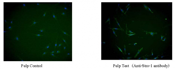
When cultured stem cells were smeared by immuno-fluorescence using antiStro-1 antibodymarker, it was found that stem cells from dental pulp had cells with Stro-1 marker expression but none from the alveolus as illustrated in picture1 and 2.
Picture 1 Stem cells from dental pulp from control group (left) and test group (right)

Pulp Control Pulp Test(Anti-Stro-1 antibody)
Picture 2 Stem cells from alveolus from control group (left) and test group (right)
Bone Control Bone Test(Anti-Stro-1 antibody)
Picture1 and 2 indicated that stem cells from dental pulp from the test group displayed green fluorescent light more than those of control group whilst stem cells from alveolus showed no difference between the two groups.
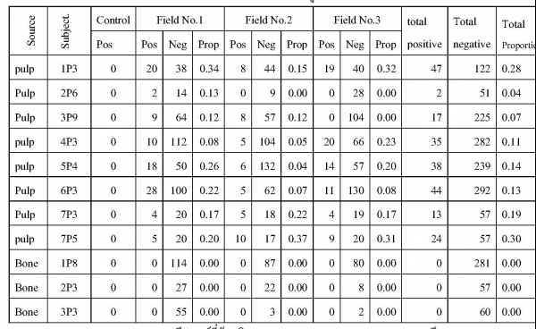
Table 1 illustrated the proportion of stem cells with Stro-1 (positive cell) to 100x total numbers of cells of dental pulp from 7 samples (8 samples) and 3 samples from alveolus.

Note: Pos = Positive cell means stem cells with Stro-1 marker.
Neg = Negative cell means stem cells with no Stro-1 marker.
Prop = Proportion means proportion of stem cells with Stro-1 marker to the total numbers of cells.
P (passage) means numbers of cell transfer (trypsinization).
According to table 1, stem cells from the dental pulp had the proportion of stem cells with Stro-1 marker to the total numbers of cells at 0.04 - 0.30 while there were no stem cells with the expression of Stro-1 marker found in the alveolus. Proportion of stem cells with Stro-1 marker to the total numbers of cells from the same sample with different passage had unequal proportion of stem cells with Stro-1 marker to the total numbers of cells as shown in sample no 7, passage#3 and 5.
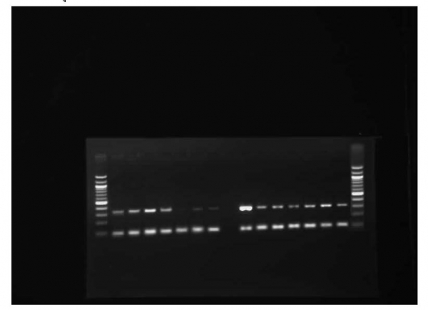
The study of The expression of CD29 and CD44 markers from reverse transcription polymerase chain reaction (RT-PCR) indicated that stem cells from both dental pulp and alveolus bore the expression of CD29 (346 bp) and mRNA GAPDH (84 bp) in every sample in which 3 in 7 of stem cells from dental pulp (vertical rows of 1 - 7) had low expression of CD29 comparing to mRNA GAPDH whilst those from alveolus (vertical rows of 8-14) had clear expression of CD29 of 7 samples as shown in picture 3.
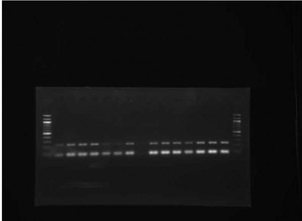
Picture 3
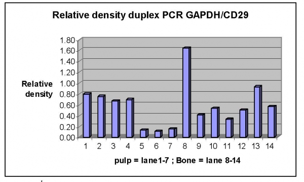
displayed the expression of CD29 in Duplex PCR. Vertical rows of 1-7 were stem cells from the tissues of dental pulp and next row was water as a negative control. Vertical rows of 8-14 were stem cells from alveolus with a 100 bp marker on the vertical left most as a control group while on the vertical right most was a test group. Upper horizontal row was CD29 (346 bp), lower horizontal row was mRNA GAPDH (84 bp) for the comparison of quantity of expression of CD29 of stem cells both from dental pulp and alveolus. Relative density was measured between CD29 and mRNA GAPDH by measuring the intensity of beam of CD29 divided by the intensity of beam of mRNA GAPDH using Scion Image26 as shown in bar graph 4.
Picture 4
bar graph illustrating relative density between CD29 and mRNA GAPDHBar graph 1 - 7 were stem cells extracted from tissues of dental pulp while bar graph 8-14 showed alveolus ones.
According to bar graph 4 it was found that stem cells from dental pulp as in sample#1-4 had relative density between 0.6-0.8 while sample# 5-7 had relative density less than 0.2. Stem cells derived from dental pulp as in sample#8 had relative density up to 1.6 while sample#9-14 relative density between 0.4-0.9. Stem cells derived from dental pulp as in sample#11 possessed the lowest relative density at 0.3.
When relative density of stem cells derived from both dental pulp and alveolus was tested by Mann-Whitney U test, no significant differences were found at reliability of .01. Therefore, the expression of CD29 from both sources was different in each sample.
The result of the expression of CD44 indicated that stem cells from both sources had the expression of CD44 (231 bp) mRNA GAPDH (84 bp) in every sample by which vertical row 5 and 6 of dental pulp had the expression of CD44 less than mRNA GAPDH while those of alveolus delivered consistent expression with no differences.

Picture 5 illustrated the expression of CD44 in Duplex PCR process.

Picture 6 Bar graph illustrating relative density between CD44 and mRNA GAPDH
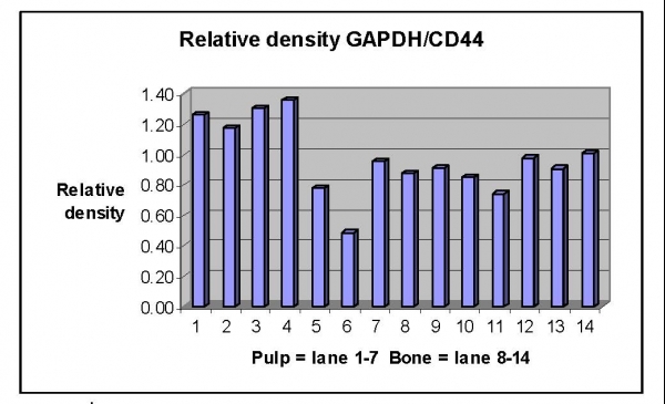
Vertical row 1 - 7 displayed stem cells generated from tissues of dental pulp and next row was water as a negative control. Vertical rows of 8-14 were stem cells from alveolus with a 100 bp marker on the vertical left most as a test group while test group was on the vertical right most. Upper horizontal row was CD29 (231 bp), lower horizontal row was mRNA GAPDH (84 bp) for the comparison of quantity of expression of CD44 of stem cells both from dental pulp and alveolus. Relative density was measured between CD44 and mRNA GAPDH by measuring the intensity of beam of CD44 divided by the intensity of beam of mRNA GAPDH using Scion Image26 as shown in bar graph 6.
Bar graph 1 - 7 were stem cells extracted from the tissues of dental pulp while bar graph 8-14 showed alveolus ones.
According to bar graph 6, it was found that stem cells from dental pulp as in sample#1-4 had relative density between 1.2 - 1.4 while sample# 5 and 6 showed the lowest relative density (0.4-0.8) of stem cells derived from dental pulp which was in accordance with the expression of CD29. However, the expression of CD44 in sample 7 was higher (relative density at 0.95) which was opposite to the expression of CD29 (relative density at 0.15) Stem cells derived from dental pulp as in sample#11 possessed relative density lower than that of other samples at 0.75. This indicated that stem cells derived from the alveolus of the sample showed no difference of the expression of CD44 which was different from those of the dental pulp.
When relative density of stem cells derived from both dental pulp and alveolus was tested by Mann-Whitney U test, no significant differences were found at the reliability of .01.
Discussion
Attributes of stem cells could be seen from the shapes of cells that were similar to fibroblastic morphology. The expression of stem cell markers, gene expression profile 27 and mesenchymal stem cell marker were divided into 3 types: positive stem cell markers such as Stro-1, CD13, CD29, CD44, CD73, CD105, CD106 and negative stem cell markers like CD11, CD31, CD34, CD45, CD117 as well as variable markers such as Sca-1, CD10, CD90 and Flk-1.28 In this study the tissues from dental pulp and alveolus were derived from mesenchymal tissues so Stro-1 marker, CD29 and CD44 were then selected for the extraction of stem cells using fluorescent smear to discover CD29 and CD44 markers.
There were various types of cells in the dental pulp. However, most of them contained columnar-shaped cells called "odontoblast" and spindle-shaped cells16 called "pulp fibroblasts". Most of cells in the bone contained osteoblast and osteocyte which had star shape.29 Cells extracted from dental pulp and alveolus in this study were fibroblastic morphology which could be fibroblast in dental pulp. Thus, the identification of stem cells was required to discover stem cells marker in which fibroblast in dental pulp had no expression of stem cells marker.30
Of the fluorescent smear using antiStro-1 antibody and the identification of stem cells by testing the expression of CD29 and CD44 with reverse transcription- polymerase chain reaction, it was found that fiboblastic morphology extracted from dental pulp showed cells with Stro-1, CD29 and CD44. It was concluded that some of extracted cells were stem cells from the dental pulp.
Of the fiboblastic morphology extracted from alveolus, there was no expression of Stro-1 found. However, the shape of cells and the identification of stem cells were conducted by reverse transcription- polymerase chain reaction, it was found that fiboblastic morphology extracted from alveolus showed the expression of CD29 and CD44 which were markers of stem cells derived from mesenchymal tissues. It was high probability that fiboblastic morphology extracted from alveolus contained stem cells.
Stro-1 was the early marker of passage when cells grew and differentiated into other types of cells. The expression of Stro-1 decreased.10
Fasai Kunthawong found that the characteristics of topography of culture dish, for instance, growth factors, hormones in the essence of Butea superba Roxb affected the expression of Stro-1 marker 31.
Shi and Gronthos applied the immulogical method using antiStro-1 antibody to smear cells from the dental pulp and tissues from bone marrow and found that smeared cells with antiStro-1 antibody would be present on the perivascular and perineural sheaths of both tissues.32
Jones and McGonagle investigated the expression of stem cells marker and found that stem cells cultured in the laboratory showed the expression of marker different from stem cells in the body in which markers that indicated stemness, for example, Stro-1 would not be discovered in the stem cells cultured in the laboratory but these cells still possessed the multipotential attribute.33
Matsubara et. al performed the experiment on the extraction of stem cells from jaw bone by piercing to draw bone marrow out of anterior ramus of the patients whose impacted tooth were operated and cyst was removed to be tested for stem cells by smearing with antiStro-1 antibody and testing for cells with the expression of CD13, CD14, CD24, CD34, CD44, CD49b, CD56, CD71, CD90, CD105, CD106, CD117, CD124, CD138 and CD144 and found that the successful rate of extraction of stem cells was 70% with the success on 29 patients out of 41.34
Papatpong Sirikururat investigated the compatibility of the stem cells from marrow and osteo- pontin/Bovine collagen using stem cells from marrow extracted from the out growth of alveolus on the palatal region that had more spongy bone than other oral regions.3
The expression of Stro-1 could not be examined in this study. It might be that the expression of Stro-1 was found on the perivascular and perineural sheaths32 but the dental pulp used as a sample of this study was taken from the out growth of the lingual arch of the lower jaw, a compact bone without blood vessels and nerves. However, stem cells could still be extracted since there might be parts of the spongy bone that might contain marrow with stem cells.
In addition, it was also discovered that stem cells with the expression of Stro-1 in the marrow were very few. Gronthos et. al drew the marrow from the iliac crest and 0.001 of total extracted cells with the expression of Stro-1 found.35
Although there was no expression of Stro-1 found in this study since stem cells were extracted from the parts of dental pulp that was cut off for the purpose of wearing denture. There should be further experiments for osteogenic induction or odontogenic induction to prove for stemness since marker expressing in the laboratory was totally different from that of the human body.33
The study of Coppe, Zhang and Den Besten indicated that stem cells from dental pulp of baby tooth displayed cells with the expression of Stro-1 at 2% of the total cells36 while the study of Shi and Gronthos32 reported that stem cells from dental pulp of the permanent tooth showed 6% of Stro-1 expressing. This study showed that the stem cells from dental pulp of permanent tooth had the expression of Stro-1 at 0.04 - 0.30%. Factors affecting the extraction of stem cells were age; perfect age range should be 20-39 years34, source of collected tissues and numbers of initial cells34, mothed of extraction16, culture condition 37, numbers of passages38 and cryopreservation.39
Samples in this study of dental pulp were female aged 17, 22, 24 and 25 years old and male aged 26, 35 and 41 years old. For alveolus samples were one female, 17 years of age and 2 male aged 27 and 68 years of age. The last sample of very old age was included since the screening process was quite difficult and might affect the expression of Stro-1 of the extracted stem cells.
Out growth method was used in this investigation. Enven it was time consuming but convenient and, above all, the small amount of extracted cells had not been damaged by enzymes as in digestion method.16 In addition, acquired cells were not single colony but contained a various types of cells that supported the growth of stem cells.10
Culture condition impacted the growth and expression of the gene of the stem cells. This study had launched alpha MEM culture media for the first year with no positive results found. DMEM was then replaced with the booster of fetal bovine serum L-glutamine, ant- fungus and anti-biotic solution in order to feed cells and prevent the infections of fungi and bacteria.
The extracted cells directly from the tissues, not cryogenized, were administered so no impacts of cryopreservation were concerned. However, the growth rate of cells could not be controlled as the out growth method was used. Thus, many passages might be conducted and the cells of generation 3-9 were used in the study. Yu et. al40 reported that stem cells from the dental pulp cultured from generation 1-9 showed the decreased proliferation rate and expression of Stro-1 conforming to this study. This study found that the proportion of cells with Stro-1 in generation 3 of stem cells was higher than that of previous generations such as generation 6-9 expcept for sample 7 of gen 5 that showed the expression of Stro-1 higher than gen 3. This was owing to the political disorder in town cuasing no passage of cells per schedule. These cells were left crowded, old and dead. They might produce some signaling factors, for instance, Growth and differentiation (GDF) and Transforming Growth factor (TGF) that stimulated the differentiation31,41,42,43 of stem cells affecting the mere generation 3 to show the proportion of Stro-1 expressing lower than that of generation 5.
CD29 (Integrin ) is transmembrane protein that mediates interactions between cells and the extracellular matrix (ECM). Apart from being marker of stem cells it was found in ovarian cancer with the property of supporting cancer cells to bind to the peritoneum resulting in metastasis.44CD44 is a cell-surface glycoprotein. It is a receptor for hyaluronic acid (HA) and adhesion molecule to the surface of tumor cells causing metastasis.44 Apart from being a marker of stem cells it is also found on the osteoprogenitor cells at the zone of hypertrophy and the zone of erosion of the bone.45
The expression of marker of stem cells was not only determined genetic46 but also by cellular circumstances and forces such as hypergravity- majority of forces specific to cells, microgravity- minority of forces specific to cells47, contractibilit)48 and shear stress.49
The expression of CD29 and CD44 in differenct samples might be from genetic factors and different mastication of each individual. The expression of CD29 had no variance to that of CD44 (Samples with low expression of CD29 may have high expression of CD44). As a result, there was no significance of the expression of both CDs between persons and between tissues from dental pulp and alveolus when statistically tested.
Periodontitis relating to genetic abnormality or inability of oral health care was found in children with Down syndrome. Using stem cells from dental pulp to build or fix or treat periodontic organs arising out of the disability from diseases might be of benefits for them. The induction of partial pulp regeneration or partial tissue of dental pulp and pulp-dentine regeneration50 were good examples. Miura et.al found that stem cells derived from a milk tooth could differentiate into 12-20 stem cells and further cultured 100% of extracted cells could form the bone.51
Nanthawan Navaroj et.al 52 conducted comparative study on anabolism of mineralized tissue from stem cells of dental pulp of normal children and children with Down syndrome and found that stem cells of children with Down syndrome showed metabolism and proliferation rate as well as created calcified tissue higher that those of normal children. It was hypothesized that milk tooth of children with Down syndrome might be collected to extract stem cells for the purpose of building bones and periodontic organs destroyed by genetic disabilities.
Conclusion
Stem cells could be extracted from both dental pulp and alveolus by using immune-fluorescence. It was found that no expression of stem cells from the out growth dental pulp at the lingual region of lower jaw was tested. But by reverse transcription polymerase chain reaction, the expression of CD29 and CD44 was found in stem cells from both dental pulp and alveolus.
This study was limited to steps of extraction and identification of stem cells. The discovery of cells with the expression of Stro-1 in smaller proportion of stem cells extracted from dental pulp or no results examined for alveolus did not mean that no stem cells. This was thanks to the overdue of early stage of stem cells since Stro-1 marker could be found only in the early stage but remained stemness. The induction of differentiation of cells, for example, building mineralized tissue and so on, proves the stemness accordingly.
st as a References1.ธีรศักดิ์ ดำรงรุ่งเรือง. สเต็มเซลล์กับการพัฒนาทางทันตกรรม. ข่าวสารทันตแพทย์ 2551;21(4):10-15.
2. กรรณิการ์ ชูเกียรติมั่น. Stem cell and dental implantation.เอกสารประกอบการประชุมวิชาการสถาบัน
ทันตกรรม กรมการแพทย์ กระทรวงสาธารณสุข. 17 กรกฎาคม2552 ณ โรงแรม มิราเคิลแกรนด์ กรุงเทพฯ : 16-50.
3. ปภาตพงศ์ ศิริคุรุรัตน์, สุพจน์ ตามสายลม,Pi-Ling Chang, สมพร สวัสดิสรรพ์.การตอบสนองของ
สโตรมัลเซลล์จากไขกระดูกมนุษย์ที่มีต่อออสทิโอพอนทินและคอลลาเจนจากผิวหนังวัวเมื่อศึกษาด้วยการเพาะเลี้ยงเซลล์. ว ทันต จุฬาฯ 2551; 31:19-32.
4. Mantesco A, Shape P. Dental stem cells for tooth regeneration and repair. Expert Opin Biol Ther 2009; 9(9):1143-54.
5. Nakao K, Morita R, Saji Y, Ishida K, Tomita Y, Ogawa M, Saitoh M, Tomooka Y, Tsuji T. The development of a bioengineered organ germ method. Nat Methods 2007;4(3):227-30.
6. Duailibi SE, Duailibi MT, Zhang W, Asrican R, Vacanti JP and Yelick PC. Bioengineered Dental Tissues Grown in the Rat jaw. J Dent Res 2008; 87(8):745-50.
7.Ikeda E, Morita R, Nakao K, Ishida K, Nakamura T, Takano-Yamamoto T, Ogawa M,
Mizuno M, Kasugai S and Tsuji T. Fully functional bioengineered tooth replacement as an organ replacement therapy. PNAS 2009; 106(32):13475-80.
8.Sonoyama W, Liu Y, Fanf D, Yamaza T, Seo BM, Zhang C, Liu H, Gronthos S, Wang CY,
Shi S, Wang S. Mesenchymal stem cell mediated functional tooth regeneration in swine. Plos One 2006; 20(1):e79.
9.Otaki S, Ueshima S, Shiraishi K, Sugiyama K, Hamada S, Yorimoto M, Matsuo O. Mesenchymal progenitor cells in adult human dental pulp and their ability to form bone when transplanted into immunocompromised mice. Cell Biol Int 2007;31(10):1191-97.
10. Feng F, Akiyama K, Liu Y, Yamaza T, Wang T-M, Chen J-H, Wang BB, Huang G T-J,
Wang S, and Shi S. Utility of PDL progenitors for in vivo tissue regeneration: a report of 3 cases. Oral Dis. 2010; 16(1): 2028.
11. Rouabhia M and Allaire P. Gingival mucosa regeneration in athymic mice using in vitro engineered human oral mucosa. Biomaterial 2010.Apr 28 [Epub ahead of print].
12. Gronthos S, Mankani m, Brahim J, Robey PG, Shi S. Postnatal human dental pulp stem cells (DPSCs) in vitro and in vivo. Proc Natl Acad Sci U S A.2000; 97(25):13625-30.
13.Iohura K, Nakashima M, Ishikawa M, Nakashima A, Akamine A. Dentine regeneration by dental pulp stem cell therapy with recombinant human bone morphogenetic protein 2.J Dent Res 2004;83 (8) : 590-95.
14. Park CH, Rios HF, Jin Q, Bland ME, Flanagan CI, Hollisten SJ and Giannobile WV. Biomimetic hybrid scaffolds for engineering human tooth ligament interfaces.
Biomaterials. 2010 Aug;31(23):5945-52. Epub 2010 May 14.15. Willumeit R, Schossig M, Clemens H, Feyeraband F. In vitro interaction of human chondrocytes and mesenchymal stem cells and of mouse macrophages with phospholipids covered metallic implant materials. Eur Cell Matter 2007; Mar 2; 13:11-25.
16. Huang G T-J, Sonoyama W, Chen J, Park SH. In vitro characterization of human dental pulp cells: various isolation methods and culturing environments. Cell Tissue Res 2006; 324:225-36.
17. Jo YY, Lee HJ, Kook SY, Chong HW, Park JY, Chung JH. Isolation and Characterization of Postnatal Stem Cells from Human Dental Tissues. Tissue Engineering 2007; 13 (04):767-73.
18. Trypsinization of adherent cells. Johns Hopkins University. GRCF: The Cell Center Rev.6/02 SOP Number 114.Available from: http://cellcenter. grcf.jhmi. edu/protocols/ trypsinizingadhercells.pdf [14 Sep 2010].
19. Donaldson JG. Immunofluorescent staining- current protocols in cell biology.Wiley online library.
Available from:http://onlinelibrary.wiley.com/doi/10.1002/0471143030.cb0403s00/pdf [14 Sep 10].20. MacArthur BD, Tare RS, Please CP, Prescott P and Oreffo ROC. A non-invasive method for in situ quantification of subpopulation behaviour in mixed culture. J.R.Soc.Interface 2006; 3:63-69.
21. Spectrophotometers BioMate 3.Operators manual. Rochester, NY,Thermo Spectronic; 2001.
22. RNA extraction kit manual.USA: Qiagen; 2002.
23. Superscript III RT-PCR.California: Invitrogen; 2003.
24. Matt Lewis. Agarose gel electrophoresis (Basic method). Available from:http://www. Methodbook . net/dna/agarogel.html [23 Sep 10].
25. Bioimaging systems. User manual. Syngene,a division of Synoptics Limited ;2000.
26.Sorensen A.Scion Image guide. Satee training. Available from:http://www.sateesite.org. [8 Oct 10].
27. Nakamura S, Yamada Y, Katagiri W, Sugito T, Ito K and Ueda M. Stem Cell Proliferation Pathways Comparison between Human Exfoliated Deciduous Teeth and Dental Pulp Stem Cells by Gene Expression Profile from Promising Dental Pulp. J Endod 2009; 1536-42.
28. Kolf CM, Cho E and Tuan RS. Biology of adult mesenchymal stem cells: regulation of niche, self renewal and differentiation. Arthritis Research&Therapy 2007; 9:204-14.
29.
Rosa AL, Crippa GE, de Oliveira PT, Taba M Jr, Lefebvre LP, Beloti MM. Human alveolar bone cell proliferation, expression of osteoblastic phenotype, and matrix mineralization on porous titanium produced by powder metallurgy. Clin Oral Implants Res. 2009; 20 (5):472-81.30.Lindroos B, Maenpaa K, Ylikomi T, Oja H, Suuronen R, Miettnen S. Characterisation of human dental stem cells and buccal mucosa fibroblasts.Biochemical and Biophysical research Communications 2008.;368:329-35.
31.
Kantawong F, Burgess KE, Jayawardena K, Hart A, Riehle MO, Oreffo RO et al. Effects of a surface topography composite with puerariae radix on human STRO-1-positive stem cells. Acta Biomater. 2010 Sep;6(9):3694-703. Epub 2010 Mar 17.32. Shi S and Gronthos S. Perivascular nich of post natal mesenchymal stem cell in human bone marrow and dental pulp. J Bone Miner Res 2003; 18:696-704.
33.
Jones E and McGonagle D. Human bone marrow mesenchymal stem cells in vivo. Rheumatology (Oxford). 2008 Feb;47(2):126-31. Epub 2007 Nov 6.34. Matsubara T, Suardita K, Ishii M, Sugiyama M, Nishimura M, Saito M et al. Alveolar Bone Marrow as a cell source for Regenerative Medicine: Differences Between Alveolar and Iliac Bone Marrow Stromal Cells. Journal of Bone and Mineral Research 2005.20(3):399-409.
35.Gronthos, S.Molecular and cellular characterization of highly purified stromal stem cells derived from human bone marrow. J Cell Sci 2003. 116(Pt 9): 1827-35.
36.
Coppe C, Zhang Y, Den Besten PK.Characterization of primary dental pulp cells in vitro. Pediatr Dent. 2009 Nov-Dec;31(7):467-71.37. Wagner W and Ho AD. Mesenchymal stem cell preparations-Comparing Apples and Oranges.Stem Cell Rev 2007; 3:239-48.
38. DAquino R, De Rosa A, Laino G, Caruso F, Guida L, Rullo R, et al.
Human dental pulp stem cells: From Biology to clinical applications. Journal of experimental zoology 2009; 312:408-15.39.